
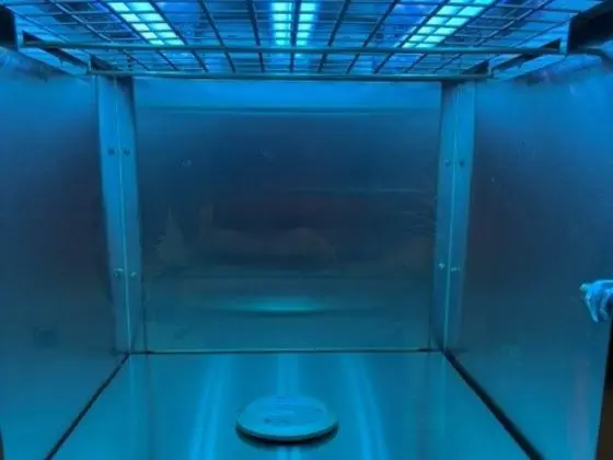
Yersinia pestis is the bacterium that causes plague, the disease behind the Black Death. It still infects people each year, mostly in parts of Africa and Asia, but also in rural areas of the United States. Germicidal UV-C light has been used against microorganisms for well over a century. A nominal dose of 26 J/m² at 254 nm is reported to inactivate more than 99% of Y. pestis.
This article explains what Yersinia pestis is, how plague spreads and what it does to the body. It then follows the history of UV in healthcare and public health, and sets out what UV-C can and cannot do.
Key takeaways
- Yersinia pestis is a Gram-negative, non-spore-forming bacterium that causes bubonic, septicaemic and pneumonic plague.
- Plague caused three major pandemics, including the 14th-century Black Death, which killed more than a third of Europe’s population.
- Today plague is treatable with antibiotics if caught early.
- A nominal UV-C dose of 26 J/m² (2.6 mJ/cm²) at 254 nm is reported to inactivate over 99% of Y. pestis.
- Germicidal UV has been used in healthcare since the 1930s, including operating theatres, TB control and hospital room disinfection.
- UV-C is a supporting control, not a cure or a replacement for medical care, pest control and hygiene.
What is Yersinia pestis?
Yersinia pestis, once known as Pasteurella pestis, is a rod-shaped, Gram-negative bacterium. It does not form spores, which matters for disinfection because vegetative bacteria are generally much easier to inactivate than spores.
Two scientists identified it during the 1894 Hong Kong outbreak: Alexandre Yersin at the Pasteur Institute and, independently, Kitasato Shibasaburō. Yersin’s name now lives on in the organism. In 1898 Paul-Louis Simond demonstrated that fleas carry the infection.
Where is it found?
The bacterium circulates in wild animals, especially burrowing rodents. Rats, mice, squirrels, prairie dogs, chipmunks, rabbits and ferrets can all host it, particularly in semi-arid grasslands and forests. Fleas pick up the bacteria by feeding on infected animals.
How does plague spread?
People usually become infected in one of three ways:
- Flea bites: the main route, via fleas that have fed on infected rodents.
- Contact with infected animals: for example, when hunters skin small animals. Pets can also bring infected fleas into the home, so cats and dogs with outdoor access can be a route in.
- Respiratory droplets: when a person with pneumonic plague coughs, others nearby can inhale the droplets. This is the only form of direct person-to-person spread.
The three forms of plague
Bubonic plague infects the lymph nodes. Typical symptoms include swollen, painful lymph nodes called buboes, fever, chills, headache and weakness. Some people develop pus-filled sores.
Pneumonic plague infects the lungs. Symptoms include high fever, headache, weakness, shortness of breath, chest pain, cough and bloody mucus. It is the most relevant form for air hygiene, because it spreads through droplets.
Septicaemic plague infects the bloodstream. Symptoms include fever, chills, weakness, nausea, vomiting, diarrhoea, abdominal pain and internal bleeding. Dark bruising of the skin can occur.
Possible complications
Without prompt treatment, plague can lead to shock, kidney failure, pneumonia and death. The World Health Organization notes that untreated bubonic plague has a high fatality rate, and pneumonic plague is almost always fatal if untreated.
Who is at higher risk?
- People living or working where rodents and fleas are common, particularly rural areas.
- People in close contact with infected people or animals.
- Laboratory workers handling cultures without suitable protective equipment.
- Hunters and others who skin or handle small wild animals.
Plague in history: Three pandemics
Ancient DNA studies have found Y. pestis in human remains thousands of years old. Historians usually describe three major pandemics:
- The Plague of Justinian (541 to 549 CE). It struck the Byzantine Empire and Mediterranean world.
- The Black Death (1346 to 1353). The deadliest outbreak, it wiped out more than a third of Europe’s population. Plague kept recurring for centuries, including the Great Plague of London in 1665.
- The Third Pandemic (mid-1800s onward). Starting in Yunnan, China, it spread worldwide through shipping routes in the 1890s and killed millions, mainly in India and China.
Before germ theory, people relied on quarantine and isolation. Antibiotics and modern healthcare later transformed outcomes.
Is plague still a threat today?
Yes, though on a very different scale. Plague remains endemic in parts of Africa, Asia and the Americas, with the Democratic Republic of the Congo, Madagascar and Peru among the most affected countries. A large 2017 outbreak in Madagascar, with many pneumonic cases, drew international attention. Rural areas of the United States also see occasional cases because of their rodent populations.
Antibiotics such as streptomycin, gentamicin, doxycycline and ciprofloxacin are effective when started early. A vaccine is available in some countries outside the US, though not in the UK or US general population. Because Y. pestis can spread by droplets and is a high-hazard pathogen, public health agencies also treat it as a biodefence priority.
Standard prevention advice
The US Centers for Disease Control and Prevention (CDC) recommends:
- Reducing rodent habitats around the home by clearing wood piles, rubbish and rocks, sealing entry points and removing exposed animal food.
- Wearing protective gloves when skinning hunted animals.
- Using flea repellent on skin and clothing outdoors.
- Treating pets regularly for fleas, taking sick pets to the vet promptly and keeping pets with outdoor access out of the bed.
- In areas with known plague, covering coughs and sneezes, washing hands regularly and informing residents of cases.
How does UV-C light inactivate bacteria and viruses?
Ultraviolet light sits between visible light and X-rays. It is split into UV-A (315 to 400 nm), UV-B (280 to 315 nm) and UV-C (100 to 280 nm). Sunlight’s UV-C is absorbed by the atmosphere, so germicidal UV-C is produced artificially.
UV-C photons are absorbed by the DNA and RNA of microorganisms. This creates molecular lesions, particularly thymine dimers, that block replication. The cell can no longer multiply and so can no longer cause infection. DNA absorbs most strongly around 260 to 265 nm, and low-pressure mercury lamps, the long-standing standard source, emit most of their energy at 254 nm. That is why reference data is usually quoted at that wavelength.
Understanding UV dose
Effectiveness depends on dose, which is irradiance multiplied by exposure time and is measured in joules per square metre (J/m²). Many older papers use mJ/cm². To convert, multiply by 10, so 1 mJ/cm² equals 10 J/m².
Different organisms need different doses. Vegetative bacteria are generally the most susceptible, enveloped viruses are also highly susceptible, and spores and some protozoan cysts need far more. Real-world conditions matter too. Shadowing, dust, organic matter, humidity, surface texture and lamp ageing can all reduce effectiveness, so practical systems are designed with safety margins above laboratory values. Some bacteria can also partly repair UV damage through photoreactivation.
What UV-C dose inactivates Yersinia pestis?
A widely cited compilation of microbial UV sensitivity data, published by the US National Institute of Standards and Technology (NIST) in 2021, lists a nominal UV-C dose of 26 J/m² (2.6 mJ/cm²) at 254 nm for better than 99% inactivation of Yersinia pestis.
| Organism | Alternate name | Type | Disease | Nominal UV-C dose for >99% inactivation |
|---|---|---|---|---|
| Yersinia pestis | Pasteurella pestis | Bacteria | Plague | 26 J/m² (2.6 mJ/cm²) at 254 nm |
Some points of context:
- A nominal dose is a laboratory reference figure. It is not a guarantee for a particular room, surface or airflow.
- Real installations are normally designed with a margin above the laboratory value, because of shadowing, organic matter, distance and lamp ageing.
- Complex or food-contact surfaces are usually evaluated at much higher doses than clean laboratory media, often in the range of thousands of J/m².
- Y. pestis is a high-hazard pathogen, so direct experimental work is carried out only in specialist containment laboratories. Systems for real-world use should be designed using peer-reviewed data and, ideally, independent validation.
It would be inappropriate to claim any UV-C product “kills plague” or prevents infection. What the evidence supports is that UV-C can inactivate this bacterium at modest laboratory doses and can be one layer within a wider control strategy.
A timeline of UV light in healthcare and public health
1877: Sunlight kills bacteria
English scientists Arthur Downes and Thomas Blunt showed that sunlight could stop bacterial growth in nutrient solutions, with the shorter, bluer wavelengths the most effective. It was the first clear demonstration that light could act as a germicide.
1903: Niels Finsen’s Nobel Prize
Danish physician Niels Finsen won the Nobel Prize in Physiology or Medicine in 1903 for treating skin tuberculosis (lupus vulgaris) with concentrated light radiation. His work helped establish phototherapy and encouraged research into ultraviolet’s biological effects.
Early 1900s: UV for drinking water
Early experiments in France led to some of the first municipal UV water-treatment installations, in Marseille in the early 1900s. Over the following century UV became one of the most widely used non-chemical methods of water disinfection, valued because it leaves no taste or chemical by-products.
1930s: Operating rooms and schools
The 1930s were a landmark decade. At Duke University, surgeon Deryl Hart installed UV lamps in operating rooms to reduce airborne bacteria and reported falling rates of post-operative infection. William and Mildred Wells in the United States studied airborne infection and the effect of UV in schools, finding less spread of measles in classrooms with upper-room UV than in those without. This research underpinned ultraviolet germicidal irradiation (UVGI) for air.
1950s to 1960s: Tuberculosis control
Tuberculosis was a leading cause of death, and UV was an attractive control. Richard Riley and colleagues in Baltimore exposed guinea pigs to air from a TB ward and showed that UV irradiation of the exhaust air substantially reduced infection. Upper-room UVGI, where lamps irradiate air near the ceiling while keeping occupants shielded, became a recognised tool. Modern US guidance from the CDC and the National Institute for Occupational Safety and Health (NIOSH) still recognises UVGI as a supplement to ventilation and filtration in TB infection control.
2000s to 2010s: Healthcare-Associated Infections
As concern grew over resistant organisms such as MRSA, Clostridioides difficile and vancomycin-resistant enterococci, hospitals began adopting mobile UV-C devices to treat patient rooms after cleaning. Some hospitals also used them during the 2014 Ebola outbreak. Studies report reduced surface contamination, though evidence on patient infection rates varies, and these devices are generally positioned as an addition to manual cleaning.
2020 Onward: COVID-19
The COVID-19 pandemic brought a surge of interest in UV for air and surface disinfection. Laboratory studies showed that SARS-CoV-2 and other coronaviruses are highly susceptible to UV-C.
Where could UV-C help against plague-type risks?
Plague control relies on rapid diagnosis, antibiotics, isolation of pneumonic cases, flea and rodent control, and protective equipment. UV-C can play a supporting role in a few situations:
- Air disinfection in rooms or ducts where droplet-borne bacteria are a concern, such as clinics or isolation areas in high-risk regions.
- Surface disinfection in laboratories, veterinary settings or facilities that handle wild animals.
- Rural and high-rodent areas, where air and surface disinfection can supplement the standard preventive measures above.
Limitations of UV-C disinfection
- Line of sight: UV-C only treats what it reaches, so shadowed areas get little or no dose.
- Surface condition: dirt, organic matter and rough materials reduce effectiveness, so clean first.
- Dose and time: insufficient exposure gives incomplete inactivation.
- Maintenance: lamps lose output with age, so monitoring and replacement schedules matter.
- Material effects: prolonged exposure can degrade some plastics and rubbers.
UV-C safety
UV-C at 254 nm can cause painful eye injury (photokeratitis) and skin erythema, so people must not be directly exposed. Safe systems use enclosures, interlocks, occupancy sensors, warning signs and trained operators, and follow relevant exposure limits. We offer free UV safety in the workplace webinar which covers the risks, exposure limits and legal requirements for working with UV.
Frequently Asked Questions
What is Yersinia pestis?
It is the Gram-negative bacterium that causes plague in humans and animals. It is usually spread by infected fleas and, in its pneumonic form, by respiratory droplets.
What UV-C dose inactivates Yersinia pestis?
A nominal dose of 26 J/m² (2.6 mJ/cm²) at 254 nm is reported to inactivate more than 99% of Y. pestis under laboratory conditions. Real-world systems are designed with a safety margin above this.
Is plague still around today?
Yes. It occurs in parts of Africa, Asia and the Americas, including rural areas of the United States. It is treatable with antibiotics if diagnosed early.
How does plague spread between people?
Only pneumonic plague spreads directly, through respiratory droplets from close contact. Bubonic and septicaemic plague usually spread through flea bites or contact with infected animals.
Does UV-C kill viruses as well as bacteria?
Yes. UV-C damages the genetic material of bacteria and viruses. Enveloped viruses such as coronaviruses are generally highly susceptible, though required doses vary by organism.
Can UV-C light prevent plague?
No single product prevents plague. UV-C can help reduce microorganisms in air and on surfaces as part of a layered strategy that also includes medical treatment, vector control and public health measures.
How do I convert UV dose from mJ/cm² to J/m²?
Multiply by 10. For example, 2.6 mJ/cm² equals 26 J/m².
Conclusion
Yersinia pestis shaped human history through three devastating pandemics and remains a risk in parts of the world. Germicidal UV has a long track record, from Downes and Blunt’s 1877 experiments and Finsen’s Nobel Prize to the operating-room, school and TB-ward studies of the 20th century, and on to COVID-19 and far-UVC research. For Y. pestis, a nominal laboratory dose of 26 J/m² at 254 nm achieves over 99% inactivation, which is why UV-C is a credible supporting measure when correctly designed, installed and used safely.
If you would like advice on whether UV-C is suitable for your application, contact our team for a consultation.
References and Further Reading
Please check each reference against the original source before publishing.
- Sensitivity of Bacteria, Protozoa, Viruses, and Other Microorganisms to Ultraviolet Radiation. Journal of Research of the National Institute of Standards and Technology, Volume 126, Article 126021 (2021).
- World Health Organization. Plague fact sheet.
- Centers for Disease Control and Prevention. Plague: How Plague Spreads; Preventing Plague; Signs and Symptoms.
- Harvard Health Publishing. Plague (Yersinia pestis).
- Encyclopaedia Britannica. Yersinia pestis.
- Antibiotic therapy of plague, National Library of Medicine (PMC8151713).
- Yersin, A. (1894). “La peste bubonique à Hong-Kong.” Annales de l’Institut Pasteur.
- Simond, P.-L. (1898). “La propagation de la peste.” Annales de l’Institut Pasteur.
- Downes, A. and Blunt, T. P. (1877). “Researches on the effect of light upon bacteria and other organisms.” Proceedings of the Royal Society of London.
- Nobel Prize Organisation. Niels Finsen: Facts.
- Wells, W. F. and Wells, M. W. (1936). “Air-borne infection.” JAMA.
- Riley, R. L. et al. (1962). “Infectiousness of air from a tuberculosis ward.” American Review of Respiratory Disease.
- NIOSH (2009). Environmental Control for Tuberculosis: Basic Upper-Room UVGI Guidelines for Healthcare Settings.
- Kowalski, W. (2009). Ultraviolet Germicidal Irradiation Handbook. Springer.
- Buonanno, M. et al. (2020). “Far-UVC light (222 nm) efficiently and safely inactivates airborne human coronaviruses.” Scientific Reports.